
Dental implants — replacing missing teeth
Dental CBCT Scans: When Is 3D Imaging Useful?
Cone-beam computed tomography (CBCT) provides three-dimensional information about teeth, jaws and surrounding structures when this additional detail is clinically justified.
Explore your tooth replacement options
If you are considering replacing a tooth or several teeth, a £30 consultation gives you a chance to explain what matters most, ask questions and explore the options with our team.
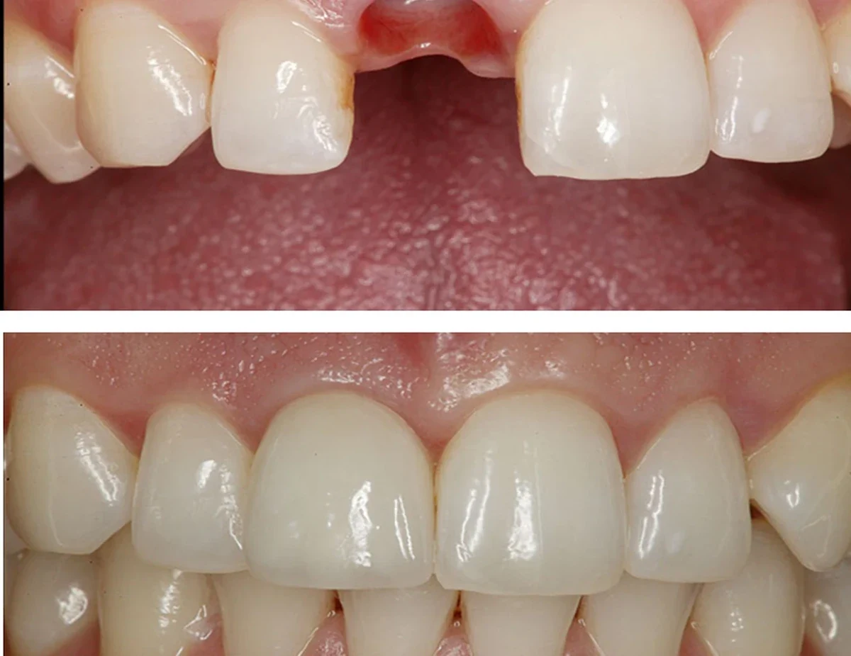
A dental CBCT scan is useful when three-dimensional information could change a particular diagnosis or treatment plan. It may help with selected implants, root-canal questions or surgery where simpler records are insufficient. The extra detail needs to justify the radiation exposure and still forms part of a clinical assessment.
What changes with three-dimensional imaging
Ordinary tooth and panoramic X-rays provide two-dimensional views, where structures may overlap. CBCT can clarify bone boundaries and the position of a proposed implant relative to the lower-jaw nerve or upper sinus. It may also help investigate selected complex anatomy that conventional imaging has not explained.
When a scan can affect a treatment decision
The justification should be linked to a clinical question: assessing bone at a proposed implant site, investigating a difficult endodontic case, evaluating impacted teeth or planning selected surgery. The scan is part of the diagnosis, not the diagnosis itself. Other findings and an examination may change the treatment options independently of the image.
Why not scan every patient?
CBCT uses ionising radiation and should only be requested where expected benefit outweighs risk, with field of view and protocol appropriate to the indication. More detailed imaging is not inherently better if the information will not change care. Existing images should be considered where suitable; unnecessary repeat exposure should be avoided.
What happens during the appointment
The equipment rotates around the head for a short acquisition while the patient keeps still. The scan may require removing relevant metallic items and following positioning instructions. A scan needs competent interpretation of the captured volume, including incidental findings requiring further attention.
What a CBCT cannot guarantee
Three-dimensional imaging can improve information available for planning but cannot eliminate surgical risk, predict exact healing or substitute for clinical judgement. It may show anatomy clearly without demonstrating whether a patient can maintain the planned prosthesis or whether a particular tooth is clinically restorable.
Each type of imaging answers a different question
A small intraoral radiograph can be excellent for examining a single tooth, root or local area of bone. A panoramic radiograph provides a broad overview of jaws and teeth. Cone-beam computed tomography (CBCT) creates three-dimensional information that can help where an implant site is close to a nerve, the sinus or a complex bony defect. None is automatically the 'best' image for every patient. Imaging should be chosen because it will change diagnosis or management; extra radiation cannot be justified merely because a practice owns sophisticated equipment.
Why an implant assessment may involve a CBCT scan
For an implant, the clinician needs to assess bone height and width and structures that must be avoided. Complex anatomy may need three-dimensional imaging, while surface scans show how the final tooth would fit the bite. Combining them depends on accurate matching and interpretation. A CBCT image alone does not demonstrate gum thickness or establish that no graft will be needed.
Interpretation matters more than the machine
Three-dimensional images can contain artefacts from metal restorations, movement or technical settings. Some findings are incidental and may require an appropriate referral or separate assessment. The dentist must examine the patient, look at the clinical context and justify the size of the scanned field. People should be able to ask why a scan is needed, which decision it will support, what the radiation exposure involves and whether the same information could be obtained through a simpler investigation.
Connect imaging to a meaningful treatment decision
The results should be explained in relation to the decision, such as comparing implant positions or investigating a tooth before root-canal care, a crown or extraction. A summary should also identify what remains uncertain, including healing and maintenance. An Iconic Smiles assessment can establish whether CBCT is indicated and how it would inform a conservative, maintainable plan.
Your next step, when you’re ready
We would be pleased to welcome you for a £30 consultation at your preferred Iconic Smiles practice. Bring your questions, and we can talk through the options that may suit you.
Any additional investigations and their fees will be explained before you decide.