
Dental implants — replacing missing teeth
Do You Need a CBCT Scan Before Dental Implants?
Cone-beam computed tomography, usually called CBCT, creates a three-dimensional view of teeth and jaw structures.
Explore your tooth replacement options
If you are considering replacing a tooth or several teeth, a £30 consultation gives you a chance to explain what matters most, ask questions and explore the options with our team.
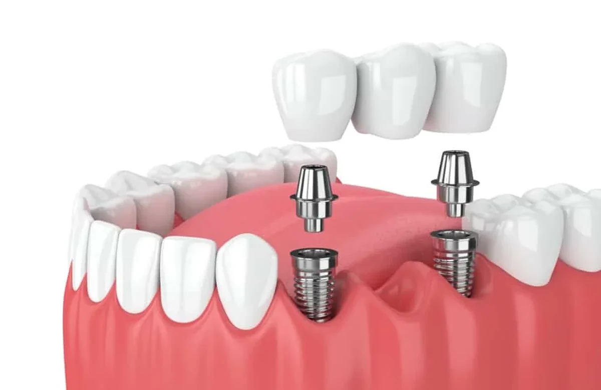
You may need a CBCT scan before an implant if three-dimensional detail would affect the surgical decision; it is not automatic for every consultation. Cone-beam computed tomography shows relationships that a flat X-ray may not define adequately. The team should explain the information needed and limit the scanned area appropriately.
What a panoramic X-ray can and cannot show
A conventional dental radiograph provides valuable information about teeth, bone height and some surrounding structures. However, the image compresses three-dimensional anatomy into two dimensions. It may not reliably show how wide a ridge is from cheek to tongue or the exact spatial relationship between the planned implant and a nearby nerve canal. An initial radiograph and clinical examination may be sufficient for some early discussions. Where precise width, orientation or anatomical boundaries would influence implant placement, a three-dimensional scan can be more informative.
The scan should answer a defined surgical question
An upper back-tooth site may need assessment of bone below the sinus, while a lower molar may be close to the nerve canal. At the front, the outer bone plate and implant angle affect where the crown can emerge. A scan should resolve a specific uncertainty in one of these relationships, with the clinician explaining how its result would change the plan.
Images cannot substitute for a restorative design
An implant must ultimately support a tooth that fits the bite, matches the planned space and can be cleaned. A three-dimensional bone image is useful only when interpreted alongside the proposed crown position and the condition of neighbouring teeth. Surface scans, photographs and diagnostic models may help establish this restorative goal. The clinician should explain whether the available bone allows the implant to be placed in a favourable position or whether grafting, orthodontic space management or a different restoration would be preferable. A technically possible implant is not always the best functional solution.
Radiation exposure needs justification
CBCT uses ionising radiation, and exposure depends on the equipment, settings and size of the scanned region. The practice should select an appropriately limited field of view and parameters that provide enough information for the clinical question. A patient does not need to avoid all useful imaging, but it is reasonable to ask why the scan is necessary and whether other available records answer the same question. Guidelines emphasise justification and optimisation rather than indiscriminate scanning. A scan ordered purely for an attractive consultation animation would not be an adequate clinical explanation.
What happens during the appointment?
The patient is positioned while the scanner rotates around the head. The exposure itself is brief, although positioning and checking the images can take longer. Metal restorations can create artefacts, and movement may affect image quality. The resulting dataset requires trained interpretation: it is not simply a photograph to be viewed by the patient. Relevant findings outside the immediate implant site may also need appropriate assessment. The dentist should explain who reviews the report and how the information is integrated into the surgical plan.
Does a CBCT scan guarantee safer implant surgery?
No imaging method removes all surgical uncertainty. CBCT can improve anatomical understanding when indicated, but the result depends on accurate interpretation, planning, surgical technique and the patient's anatomy. If guided surgery is proposed, the scan may form part of the guide design, yet guide fit and intraoperative checks remain necessary. The patient should receive a written explanation of the planned implant position, any bone augmentation and the remaining clinical uncertainties. Good imaging supports informed decision-making; it is not a standalone guarantee of outcome.
References and further reading
Book an assessment
The practices are in Hatfield and Cheshunt in Hertfordshire, and Friern Barnet in North London. For travel from Bedfordshire, Buckinghamshire or Essex, confirm the actual practice and any sites involved in later stages.
Pandya (GDC 114822).Content updated: 26 October 2026.
CBCT Before Dental Implants: The Questions a 3D Scan Answers
Three-dimensional imaging is useful when it could change care. The scan should have a specific question to answer, rather than be included automatically in every implant assessment.
A lower molar space looks adequate on a panoramic radiograph but the canal and lingual concavity cannot be mapped confidently.
A limited-area CBCT may clarify the lower-jaw nerve canal or a variation called an anterior loop where a panoramic view is insufficient. It may also help assess the hollow on the tongue side of the jaw. Planning still needs to account for drill and implant dimensions, the intended crown's emergence and any image artefacts. A scan based on an unsuitable replacement-tooth plan can mislead.
The explanation should cover radiation exposure, nerve proximity, cheek-to-tongue bone width and any relevant sinus boundary. Computer outlines of the anatomy assist interpretation but cannot establish how tissues will heal or remove surgical uncertainty.
Your next step, when you’re ready
We would be pleased to welcome you for a £30 consultation at your preferred Iconic Smiles practice. Bring your questions, and we can talk through the options that may suit you.
Any additional investigations and their fees will be explained before you decide.