
Repairing your teeth
Dental Before-and-After Photos: What Can They Show?
Dental before-and-after photographs can help illustrate visible changes, but they cannot alone establish treatment suitability, function, long-term survival or the experience of another patient.
Talk through your options for repairing teeth
If you would like to understand how a tooth might be repaired or protected, a £30 consultation offers time to discuss your concerns and what an individual assessment may involve.
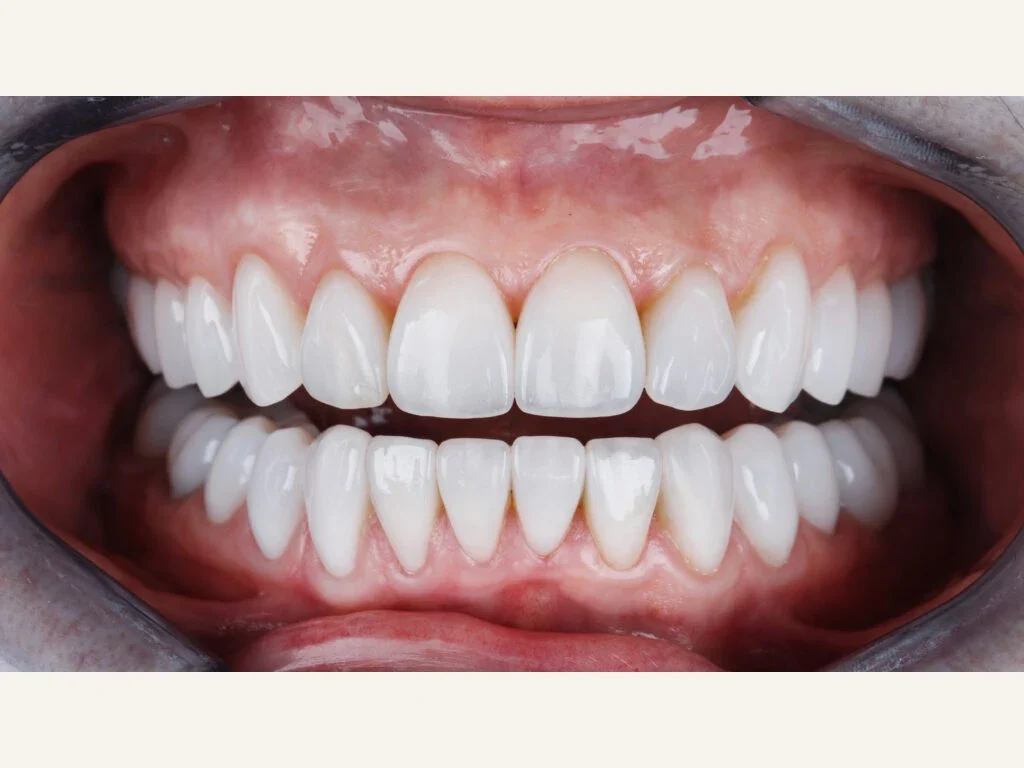
Before-and-after photographs can help you compare visible changes in tooth shape, position or colour. They cannot show everything about the bite, health, maintenance or long-term result, and another person's outcome cannot predict yours. Look for context about the treatment and clear labels distinguishing completed care from a simulation.
What a photograph can show
Comparable images may demonstrate selected changes in visible tooth shape, alignment, colour or soft tissue appearance at recorded times. They can support an explanation of the procedures performed. They do not reveal how an implant integrated, whether gum disease remained stable or the patient’s full medical and dental circumstances.
Why standardisation matters
Different exposure, white balance, camera distance, moisture, smiles, lip retraction and framing can exaggerate or obscure change. Clinical comparisons should use consistent acquisition methods where possible and label the treatment stage. Whitening and restoration shade look different under different light; a phone filter is not a legitimate clinical outcome measure.
Consent and privacy are essential
Consent to photographs for a clinical record is separate from permission to publish them for marketing. Identifying features, image metadata and case details need careful handling. A published result should belong to the patient described; stock images or generated faces should never be presented as evidence of someone's treatment.
Simulations must be labelled
Digital smile previews, wax-ups and AI-generated illustrations are planning tools, not photographs of completed care. Labelling them visibly avoids confusing possibility with demonstrated outcome. An unrealistically perfect rendering can create expectations that ignore anatomy, material and biological limits.
What case information helps patients
A useful case explanation sets out the original concern, important findings, alternatives, chosen treatment and the stage shown. It should make relevant limits clear. Where authorised follow-up information is available, its date helps distinguish a later result from one photographed immediately after treatment. A single example cannot guarantee the same outcome for everyone.
How to use images wisely
Use images to start a discussion about your own options. The dentist can explain whether a similar approach is appropriate, which preparation might be needed and what risks and maintenance it would involve. Your teeth and health need an independent assessment before a photographed look becomes a treatment goal.
A photograph documents appearance, not the whole outcome
Before-and-after photographs can help a patient understand possible changes in tooth arrangement, shape and shade, but lighting, head position, lip retraction and camera settings can alter perception. An impressive smile photograph does not show whether gum disease was controlled, whether an implant can be cleaned or how much enamel was removed. Nor does one person's result establish what a different bite, bone anatomy or medical history will permit.
Meaningful cases should describe the treatment, its duration where verified and any relevant qualifications about the images. Digital mock-ups and simulated smiles need clear labelling because they depict proposals rather than completed outcomes. Patients should be able to ask which parts of the result were achieved through orthodontics, whitening, bonding, veneers or replacement teeth, and what long-term maintenance the case requires.
When choosing a practice, compare more than photographs: examine the written plan, the explanation of alternatives, the clinician's relevant qualifications and the route to review or repair. Personal images are health information, and patients should know how consent for public use was obtained and how it can be withdrawn within applicable arrangements.
Treat photographs as supporting evidence, not a promise
A before-and-after pair may fail to show how the patient bites, speaks or cleans around restorations. The final photograph may be taken immediately after bonding, before any wear, staining or maintenance becomes apparent. Ask whether the images show the same patient, under comparable lighting and camera settings, and whether the treatment included orthodontics, whitening or other steps not visible in the caption.
If your own photographs are taken, you should know how identifiable images will be stored, who can access them and the arrangements for consent or withdrawal of public use. When viewing other people's results, photographs are useful alongside an explanation of clinical limits and continuing care.
Sources and professional guidance
Applying the information:
Your next step, when you’re ready
We would be pleased to welcome you for a £30 consultation at your preferred Iconic Smiles practice. Bring your questions, and we can talk through the options that may suit you.
Any additional investigations and their fees will be explained before you decide.