
Repairing your teeth
Dental X-Rays Explained: Types, Safety and When They Are Necessary
Dental radiographs help investigate disease or anatomy that cannot be assessed reliably by looking at the visible tooth surfaces.
Talk through your options for repairing teeth
If you would like to understand how a tooth might be repaired or protected, a £30 consultation offers time to discuss your concerns and what an individual assessment may involve.
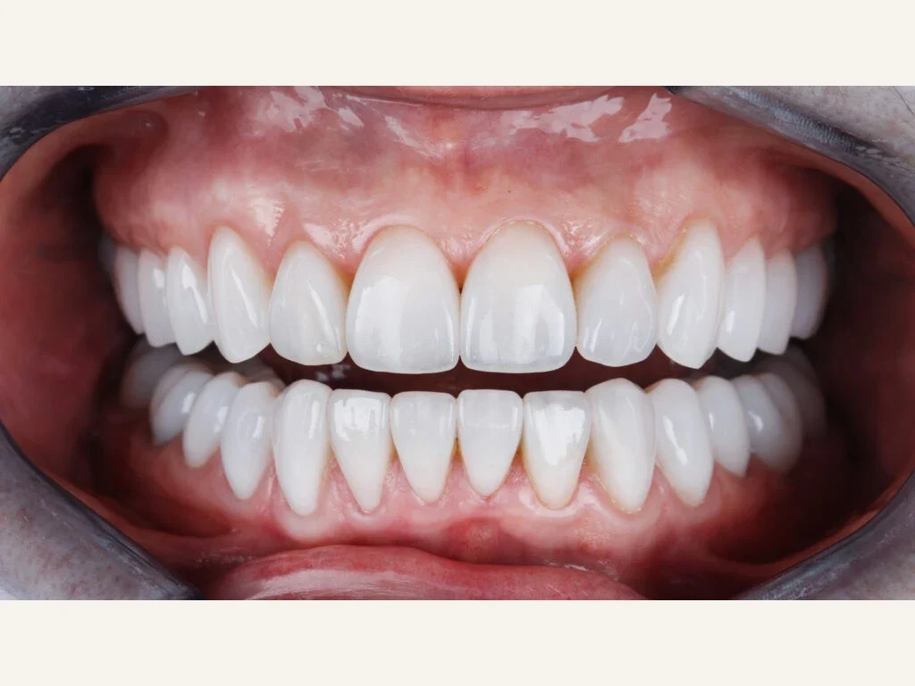
Dental radiographs help investigate disease or anatomy that cannot be assessed reliably by looking at the visible tooth surfaces. Bitewings, periapical views, panoramic images and CBCT answer different questions. Imaging should be selected and justified for the individual situation, not taken at fixed intervals without considering risk.
What common X-ray types show
Bitewing X-rays help assess decay between teeth and parts of the bone support. A periapical view shows a tooth, its root and the surrounding area; a panoramic image gives a wider view of teeth and jaws. Cone-beam CT, or CBCT, adds three-dimensional information for selected questions. The most complex scan is not necessarily the appropriate one for a straightforward concern.
How the decision is made
Symptoms, previous disease, age, clinical findings and earlier images influence whether new radiographs are indicated. A low-risk patient with recent adequate imaging may not need the same frequency as someone with active disease or unexplained symptoms. The practitioner should be able to explain what information is expected and how it could change management.
Understanding radiation safety
Dental radiography involves controlled ionising radiation. Equipment maintenance, trained operators, appropriate exposure settings and a clinically justified field help balance diagnostic benefit with radiation risk. A precise exposure comparison depends on the type and settings of the examination; blanket claims that all dental imaging carries zero risk are incorrect.
What images cannot tell us
An X-ray complements but does not substitute for a clinical examination, dental sensibility testing, periodontal measurements or a detailed history. Some cracks and early disease may not appear clearly, and an image can also contain artefacts. A normal-looking radiograph does not automatically explain away persistent pain.
Special circumstances
If you are pregnant or think you may be pregnant, tell the dental team so the indication and practical arrangements can be considered properly. Necessary care should not be automatically abandoned without discussing benefits and risks. Where previous imaging is available, ask whether it can help avoid unnecessary repeats.
A practical patient checklist
Before imaging, understand what the image is intended to detect, why it is useful now and whether another test could answer the question. The team should explain when results will be discussed and which decision they inform, alongside the fee.
Why a CBCT scan is not automatically better than a conventional X-ray
A cone-beam CT image can show three-dimensional relationships, which may assist when planning particular implant sites, assessing complex anatomy or investigating a concern not resolved by suitable two-dimensional imaging. It generally involves a different radiation exposure and creates a larger volume of anatomical information than a single intraoral radiograph. That is not a reason to use it for every dental check-up. The clinician should identify the diagnostic question, assess whether a lower-dose method can answer it, select the appropriate field of view and ensure images are interpreted competently.
What patients can ask about safety
Radiation decisions consider the expected clinical benefit against the exposure involved; comparisons between individual scans need care because settings and equipment vary. Ask what change in diagnosis or treatment is anticipated from the image and whether previous suitable scans are available. There is no clinical benefit in repeating an image simply because a practice owns newer equipment. The important standard is justified, optimised imaging with appropriate reporting and storage, not the marketing term ‘3D’ alone.
For routine imaging, the interval between X-rays should be determined by clinical findings and risk, rather than a fixed calendar rule for everyone. A patient with a history of frequent interproximal decay or new symptoms may have different imaging needs from someone whose disease has remained stable. It is reasonable to ask for the image to be explained at the chairside: where is the suspected lesion, what else might account for the symptoms and would the recommended treatment differ if no image were taken? When dental records are transferred between practices, relevant previous radiographs can provide a useful comparison and may avoid an unnecessary repeat exposure.
Sources and professional guidance
Applying the information:
Treatment options · Our practices · Fees and finance
Bitewing X-Rays: What They Add to a Dental Examination
Bitewings can show decay between back teeth and aspects of the supporting bone that direct inspection misses. Their use needs to be justified by your risk and clinical findings.
Clinical assessment and suitability
Symptoms, medical history, examination findings and earlier images help establish what a new X-ray could add. The examination and image answer different questions; neither should be expected to replace the other.
Depending on the findings, the next step may be monitoring, suitable imaging, a focused referral or treatment supported by the diagnosis and your preferences. Previous image quality, current risk and what can already be seen clinically matter. Photographs cannot show all the areas visible on a bitewing.
Repeat bitewings should follow risk and the usefulness of previous images, rather than an automatic annual schedule. Even with little recent decay, a new image may be appropriate for a particular finding. The dentist should explain how it could affect care.
One image can miss disease or give a misleading impression if an isolated sign is overinterpreted. Unnecessary imaging adds exposure without benefit, while suspicious soft-tissue changes still need appropriate assessment or referral. The image should be considered with the wider findings.
Your next step
Areas we serve: Hertfordshire · Bedfordshire · Buckinghamshire · Essex. Our actual clinics are in Hatfield, Cheshunt and Friern Barnet; Bedfordshire, Buckinghamshire and Essex are patient catchment areas, not additional clinic sites.
AI-Assisted Dental X-Rays: A Second Reader, Not a Diagnosis
AI software may highlight an area for the dentist to examine more closely. A marked pixel or confidence score cannot establish disease without comparison with the clinical findings.
How the dentist checks a software suggestion
The clinician considers image quality, earlier X-rays, symptoms and the examination, including the possibility of a false positive. Digital or conventional records, selected further imaging or referral may help when needed; extra scans may add nothing if the question is already answered. Responsibility for the diagnosis remains with the treating clinician.
A software-marked shadow may come from overlapping anatomy or image noise rather than decay. The dentist relates it to the tooth's appearance, symptoms and earlier records before recommending treatment. Automated highlighting assists that review without replacing it.
If treatment is agreed after diagnosis, the restoration should remain accessible for later examination. Its margins, retention, contacts and surrounding tissues need to be checked as part of continuing care.
Research and guidance
- NHS: dental treatment information — background reading
Your next step, when you’re ready
We would be pleased to welcome you for a £30 consultation at your preferred Iconic Smiles practice. Bring your questions, and we can talk through the options that may suit you.
Any additional investigations and their fees will be explained before you decide.