Repairing your teeth
3D Digital Planning for Complex Dental Treatment
Three-dimensional planning can combine appropriate scans, digital models and restorative design to explore treatment options before irreversible procedures.
Talk through your options for repairing teeth
If you would like to understand how a tooth might be repaired or protected, a £30 consultation offers time to discuss your concerns and what an individual assessment may involve.
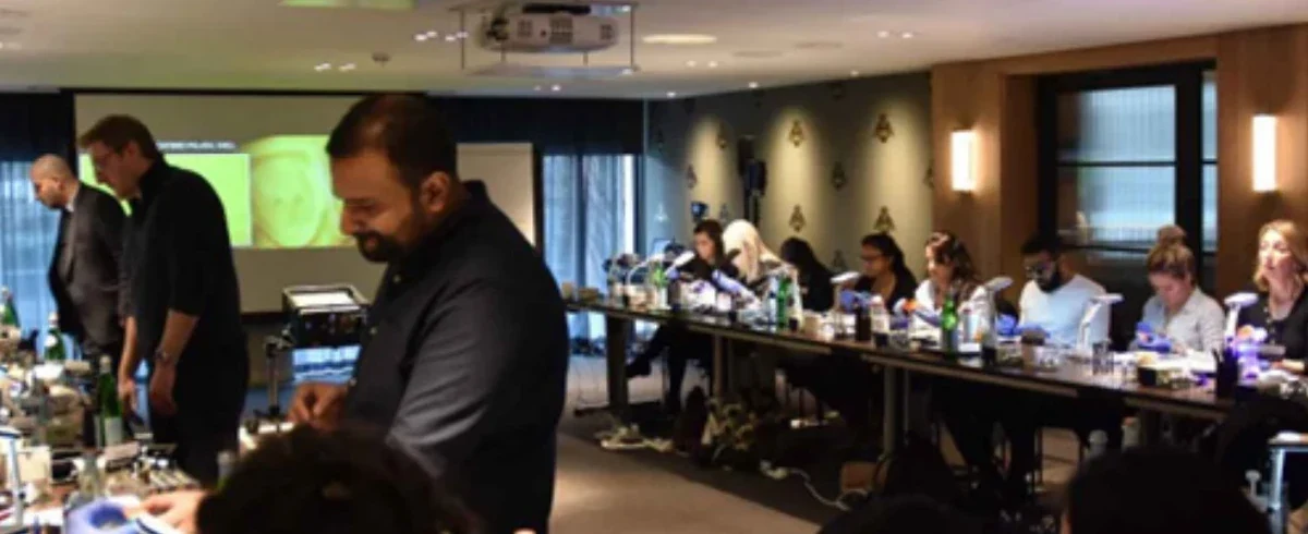
3D planning brings suitable scans, digital models and the intended tooth design together so you can explore treatment before irreversible steps. It helps the team assess space, sequence and alternatives. The image is still a plan: it cannot guarantee surgical accuracy, tooth movement or the exact final appearance.
Begin with the treatment objective
Planning should start with the desired functional and maintainable result: tooth position, support, gum health, bite and cleaning access. The clinician then decides which records are necessary. A beautiful simulation created before diagnosing active decay or gum disease risks prioritising appearance ahead of the condition that must support the result.
Combining records carefully
A surface scan records tooth contours, photographs add appearance and context, and indicated X-rays or CBCT provide different anatomical information. Matching these records can guide implant position, restorative space and the order of orthodontic care. Each record has limits, and errors in capturing or matching them can affect later stages. Clinical checks remain necessary as the plan is put into practice.
From virtual design to physical treatment
The design may be used to make a surgical guide, trial shape, printed model or final restoration. These have manufacturing limits, and a guide must seat accurately in the mouth. The clinician still checks the anatomy and responds if conditions differ from the model. Software recommendations support, rather than replace, diagnosis and responsibility for care.
Why complex work is staged
Periodontal stabilisation, tooth preservation, orthodontic movement, surgery and restoration may depend on one another. Digital planning helps show sequence and possible alternatives, including temporary stages that preserve function. Treatment times and final shapes can change when healing, response or anatomy differs from the original model.
Communicating realistic expectations
Patients can use digital visuals to understand proportion, bite contacts and likely aesthetic direction. Images should be labelled as planning simulations rather than guaranteed outcomes. A predicted implant trajectory or animated smile must not obscure the need for explicit discussion of risks, costs, maintenance and non-treatment options.
What distinguishes a responsible workflow
A useful explanation shows which records informed the plan, what has been checked clinically and what remains uncertain. You should also know how changes would be discussed. The value lies in how the information helps a particular decision, rather than the size of the screen or the equipment's price.
A digital plan is a model, not the treatment itself
Computer-guided planning can help clinicians compare proposed tooth shapes, assess space or visualise relationships between a restorative design and the underlying anatomy. In implant care, a plan may combine a surface scan and appropriately indicated radiographic imaging; each data set has different limitations. Errors in matching scans, inaccuracies in the surface record or the way a guide is used can affect translation from computer screen to mouth.
For orthodontics, staged tooth movement is predicted by software but achieved movement may differ because of biology, appliance fit and patient wear. A digital smile simulation can help discuss visible proportions without establishing how a gingival margin or final bite will behave. The clinician remains responsible for examination, risk assessment, informed consent and modification of the plan when conditions change.
The useful question is therefore not whether the practice has a 3D screen but how the data influence a specific decision. Ask what is measured, which assumptions may fail, how the final work is checked and what happens if the outcome differs from the proposal.
Identify where errors can enter a digital workflow
Digital planning involves separate steps: recording the patient's anatomy, aligning scans, defining the proposed result, manufacturing components and delivering the procedure. In implant surgery, an apparently precise virtual implant position may be affected by the stability of a surgical guide, the accuracy of registration and movement during surgery. In orthodontics, the predicted position of a tooth can differ from biological movement because appliances do not always achieve every programmed movement.
A responsible plan specifies when a new scan, X-ray or clinical examination is needed to reassess progress. Patients can ask who approves the plan, how discrepancies are managed and whether the final restoration will remain cleanable. A scan should be performed because the answer is likely to influence treatment, not simply because the equipment is available.
Sources and professional guidance
Applying the information:
Treatment options · Our practices · Fees and finance
3D Printed Dental Models in Smile and Implant Planning
A printed model is only as reliable as the scan and print calibration; it can help demonstrate contours but does not confirm tissue biology.
Alternatives and clinical trade-offs
Planning may use digital records, conventional records, a combination or referral for particular imaging. Extra scans may be unnecessary where the examination already answers the question. For a printed model, scale, surface detail, distortion and fit need checks before it informs a clinical decision.
A printed trial can help you judge tooth dimensions and how you respond to a proposed shape. Its usefulness depends on accurate measurements and printer calibration. Rigid resin cannot reproduce soft-tissue changes or predict biological healing, so its fit and appearance still need clinical interpretation.
Further reading (background sources)
- — background reading
- — background reading
- NHS: dental treatment information — background reading
Your next step
Areas we serve: Hertfordshire · Bedfordshire · Buckinghamshire · Essex. Our actual clinics are in Hatfield, Cheshunt and Friern Barnet; Bedfordshire, Buckinghamshire and Essex are patient catchment areas, not additional clinic sites.
Further questions to consider before treatment
- Digital and conventional impressions both serve planning needs. The team can explain which suits your treatment and whether both are needed.
- Scans may support crowns, implants or aligners; understand which decisions they inform and how they affect the proposed visits.
- Bite records help clinicians understand how teeth meet before planning multiple restorations.
- Digital design and manufacture can help shape a crown, with clinical planning and checks of its fit still essential.
- A digital bite simulation helps planning, while an examination checks how the teeth actually meet.
Further clinical details: How digital planning helps when several teeth need repairing
Digital planning can help coordinate care when several teeth are missing, worn or heavily restored and one repair would not address the whole situation. Scans, photographs, models and laboratory designs show how proposed stages relate to one another. Their value is in supporting a clear plan for tooth preservation, gums, bite, appearance and maintenance.
Start with a complete diagnosis, not a digital smile picture
A patient with worn front teeth and old crowns may also have changing gum levels, missing back teeth or unbalanced chewing contacts. The dentist should examine individual tooth prognosis, periodontal support, relevant radiographs, existing root treatments and functional relationships between the jaws. A digital scan records tooth surfaces and can show visible wear, but it does not independently diagnose disease beneath restorations or measure all supporting bone. The clinical team must decide which teeth are healthy enough to retain and which problems should be stabilised before definitive cosmetic design.
Decide whether the bite needs changing
Some patients can be restored while preserving their habitual jaw relationship. Others have lost so much tooth structure that a carefully planned alteration in vertical dimension or tooth contacts is considered. That decision may affect speech, muscle comfort, the position of front edges and the space available for ceramic restorations. Computer software can illustrate a proposed shape, yet it cannot guarantee adaptation to a new bite. Clinicians may use mock-ups, provisional restorations or staged additions to test certain changes. The patient should understand which aspects of the bite are being altered and why.
Treatment staging protects decision-making
Elective reconstruction should not begin with porcelain on teeth that have untreated decay or active periodontal inflammation. Stabilisation may involve hygiene, periodontal therapy, restoring salvageable teeth, treating disease or reviewing medical factors. Later stages might include orthodontics to create space, implant planning for missing teeth, and definitive restorations. Not every patient needs all these disciplines. A multidisciplinary plan is valuable when it shows how each stage supports the next and identifies which findings could change the sequence. The clinical team should avoid committing to an irreversible aesthetic design before the foundations are understood.
Digital records help different clinicians coordinate
Scans, suitable photographs and models help technicians and clinicians share the proposed tooth sizes and contours. Where CBCT is justified for an implant, it adds different information about the underlying anatomy. These records need to be interpreted with the examination. You should know who coordinates the final restoration and how differences between surgical and restorative proposals would be resolved.
Material choices vary from tooth to tooth
A heavily restored molar and a healthy front incisor need not receive the same type of treatment. Some teeth may benefit from direct composite, others from partial-coverage ceramic, and some from crowns or implant-supported replacements. Using identical materials across the entire mouth can be convenient for marketing but may remove unnecessary tooth structure. The dentist should explain why each tooth is being covered, preserved or left untouched. The selected material must fit the bite and cleaning requirements as well as the desired shade. Maintenance planning should begin before the final laboratory order.
Previewing a new smile has limits
Digital mock-ups are excellent discussion tools for tooth proportions, symmetry and relative edge positions. They can help the patient decide whether the desired change is subtle or conspicuous. However, a rendering cannot predict soft-tissue response, exact ceramic colour in every light or future biological change. A trial smile or provisional phase may provide more useful real-world feedback where clinically feasible. The patient should know which parts of the final design are flexible and which have already been constrained by anatomy or preparation.
Ask for a phased plan and an honest total estimate
The written proposal should identify diagnoses, teeth involved, stages, temporary work, visits, laboratory requirements and maintenance responsibilities. Separate essential disease control from elective changes and explain possible additional care before consent. If treatment spans months, include eating and appearance between stages. A successful rehabilitation needs these practical arrangements alongside coordinated function and appearance.
References and further reading
Book an assessment
The practices are in Hatfield and Cheshunt in Hertfordshire, and Friern Barnet in North London. For travel from Bedfordshire, Buckinghamshire or Essex, confirm the actual practice and any sites involved in later stages.
Editorially updated: 27 October 2026.
Your next step, when you’re ready
We would be pleased to welcome you for a £30 consultation at your preferred Iconic Smiles practice. Bring your questions, and we can talk through the options that may suit you.
Any additional investigations and their fees will be explained before you decide.